
The Immunohistochemistry and Immunocytochemistry techniques use antibodies (remember this entry on Antibodies for Immunohistochemistry ) to detect and visualize the presence, distribution and location of a certain protein. Although they are less sensitive immunoassays than a Western Blot or an ELISA can be , they allow the observation of processes in the context of intact cells and tissues.
Although, sometimes, the terms are used interchangeably to refer to either of the two techniques, in this post we detail the main differences between Immunohistochemistry and Immunocytochemistry.
Differences Between Immunohistochemistry And Immunocytochemistry
1.- Sample Type: Tissue Vs Cells
As its name suggests, immunohistochemistry (IHC) uses tissue sections (paraffin embedded or frozen), while immunocytochemistry (ICC) is applied to intact cells (isolated or in culture).
While immunocytochemistry (ICC) is carried out on samples of intact cells that have had all or most of the extracellular matrix removed, immunohistochemistry (IHC) is performed on samples of biological tissue where the cells are surrounded of the corresponding tissue architecture and of other cells present in it.
2.- Sample Source
In the case of Immunohistochemistry (IHC), the tissues obtained directly from the patients are preserved by freezing or by soaking them in paraffin, to subsequently section them and proceed to staining with the antibodies. In this way, the location of the cellular components of interest can be visualized while keeping the surrounding tissue architecture intact.
In immunocytochemistry (ICC), virtually all of the extracellular matrix and other stromal components are removed, leaving only whole cells for staining. The cells can be obtained from suspension samples (aspirates, blood smears, swabs …) or from cell cultures obtained in the laboratory.
3.- Sample Processing
In addition to the source from which samples are obtained, immunohistochemistry (IHC) and immunocytochemistry (ICC) also differ in their processing. Some of the main differences are:
- PRESERVATION OF SAMPLES
In the case of immunohistochemistry (IHC), the samples are frozen or paraffin-embedded to preserve tissue morphology. This step does not occur in immunocytochemistry (ICC).
- FIXATION
In immunohistochemistry (IHC), the use of binding reagents such as formaldehyde, can mask certain epitopes, limiting antigen-antibody binding. To avoid this, it is common to use antigen recovery methods to reestablish tissue antigenicity, which is not necessary in the case of immunocytochemistry (ICC).
Regarding the fixation period to which the samples are subjected, it is less in the case of immunocytochemistry (ICC) than in immunohistochemistry (IHC).
- PERMEABILIZATION
In immunocytochemistry (ICC), the cells must be permeabilized so that the antibodies can access the intracellular targets. However, in the case of immunohistochemistry (IHC), depending on the thickness of the sections and the method of fixation, the samples may not need additional permeabilization steps.
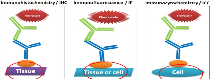
4.- Marking Method: Chromogenic Vs Fluorescent
Antigen-antibody interaction can be visualized by chromogenic detection, where an enzyme conjugated to an antibody reacts with a substrate giving rise to a color precipitate, or by fluorescent detection, where the antibody is conjugated to a fluorophore that will be visible under a microscope. fluorescence.
Although traditionally the detection method in both immunohistochemistry (IHC) and immunocytochemistry (ICC) has been through the use of chromogenic reagents, the use of immunofluorescence (IF) is increasingly widespread, especially in the case of immunocytochemistry ( ICC / IF), and increasingly in immunohistochemistry (fluorescent IHC).
Although the terms are used interchangeably on many occasions, we hope that this entry has helped you to understand the main differences between immunohistochemistry and immunocytochemistry.